GHK-Cu Basics: A Copper-Binding Tripeptide With an Endogenous Role
GHK-Cu is not a fully synthetic construct. The tripeptide glycyl-L-histidyl-L-lysine (GHK) occurs naturally in human plasma, saliva, and urine, and its plasma concentration declines with age, from roughly 200 nanograms per millilitre in young adults to around 80 nanograms per millilitre in older individuals. Loren Pickart's discovery of GHK in 1973 came during work on plasma factors that cause ageing hepatocytes to recover synthetic behaviour characteristic of younger cells: the tripeptide was isolated as the active molecule responsible for this effect.
The copper-complexed form, GHK-Cu, forms when the tripeptide coordinates a Cu(II) ion through the imidazole nitrogen of histidine and the N-terminal amine, creating a stable chelate that delivers copper to target enzymes without releasing free ionic copper (a distinction relevant to safety). Free Cu²+ is pro-oxidant and cytotoxic at elevated concentrations; the chelated form delivers copper's enzyme-cofactor benefits while limiting its oxidative liabilities. In the UK, GHK-Cu appears on product labels under the INCI name Copper Tripeptide-1 and is a permitted cosmetic ingredient without specific concentration limits.
The 4,000-Gene Claim: What the Pickart Microarray Actually Measured
The headline figure associated with GHK-Cu in much of the research literature (that it modulates more than 4,000 genes in human fibroblasts) originates from microarray studies conducted by Pickart, Vasquez-Soltero, and Margolina and published in 2010. The analysis identified genes upregulated or downregulated by more than 50% following GHK-Cu exposure in cultured human dermal fibroblasts.
The breadth of this transcriptional response is genuine and noteworthy. Upregulated networks included DNA-repair genes, antioxidant defence systems, and tissue-remodelling proteases alongside their inhibitors. What researchers must keep in mind is what microarray data do represent and what it does not. A 50% change in transcript level measured by microarray correlation is not the same as a 50% change in protein output, and protein output change is not the same as functional biological change in a tissue context. The microarray finding establishes that GHK-Cu has substantial and coordinated transcriptional effects at the concentrations tested — it does not independently establish that each of the 4,000-plus gene changes translates to a discrete biological outcome.
The detailed evidence review of GHK-Cu in dermal regeneration research situates the microarray findings within the broader evidence architecture.
Strongest Signals: Collagen Synthesis, Anti-Fibrosis, and MMP/TIMP Balance
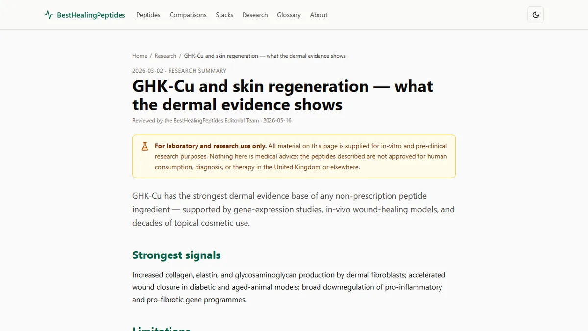
The most reproducible and mechanistically grounded findings in GHK-Cu research concern collagen synthesis in dermal fibroblasts. Multiple independent studies (including Maquart and colleagues in 1997) have demonstrated that nanomolar concentrations of GHK-Cu stimulate type I and type III collagen mRNA expression and secretion, with a defined dose-response relationship and parallel induction of glycosaminoglycans and the proteoglycan decorin. This is not a single-laboratory finding; the collagen-stimulation effect is among the best-replicated observations in the GHK-Cu literature.
The anti-fibrotic effect is a more nuanced and arguably more clinically significant observation. Many collagen-stimulating compounds simultaneously increase fibrotic scar formation. GHK-Cu appears to promote organised, mature collagen deposition while suppressing TGF-beta-1-driven myofibroblast differentiation (the cellular event responsible for pathological scarring). Animal models of pulmonary fibrosis induced by bleomycin show reduced fibrotic markers and lower collagen deposition scores in GHK-Cu-treated groups compared with controls. The capacity to increase collagen quality without increasing fibrosis risk is a genuinely distinctive pharmacological feature.
The MMP and TIMP balance (matrix metalloproteinases that degrade damaged collagen and their tissue inhibitors) is also meaningfully modulated. GHK-Cu upregulates both enzymes and inhibitors in a pattern consistent with coordinated remodelling rather than either pure matrix breakdown or pure matrix accumulation. This distinguishes the transcriptional profile from what would be observed with an uncoupled collagenase activator or a pure synthetic stimulus.
Evidence in Wound Models: Diabetic Skin, Aged-Animal Studies, and Burn Research
In wound models, the GHK-Cu evidence extends beyond cell culture. Arul and colleagues in 2012 conducted topical GHK-Cu experiments in streptozotocin-induced diabetic rats, a model of impaired healing where angiogenesis, epithelial migration, and collagen synthesis are all compromised. Animals treated with GHK-Cu hydrogels showed significantly accelerated full-thickness wound closure compared with untreated controls, alongside histological evidence of increased granulation tissue and higher hydroxyproline content (a biochemical marker for collagen deposition).
Aged-animal studies explore a different dimension of the same biology: whether GHK-Cu can restore the declining wound-repair capacity that correlates with its falling plasma concentration. Evidence in this area is promising but less extensively replicated. Burn research adds a third context in which GHK-Cu's anti-fibrotic properties are particularly relevant, given that hypertrophic scar formation is a primary morbidity of severe burns.
Taken together, these wound model findings support the cell-culture mechanism: GHK-Cu promotes collagen deposition and organisation in compromised healing contexts. What they do not establish is the dose-response relationship in human skin, the optimal formulation vehicle, or the relative contribution of different mechanisms at physiological versus pharmacological concentrations.
Where the Evidence Thins: Clinical Trials, Concentration-Response Nonlinearity
The GHK-Cu literature thins considerably at the human clinical trial level. ProCyte Corporation conducted wound-care studies in the late 1980s and early 1990s that produced commercially interesting but ultimately unpursued results. Hair-follicle research in clinical populations is limited and generally small. The cosmetic dermatology market has funded the broadest human exposure to topical GHK-Cu, but cosmetic studies are not typically powered for mechanistic endpoints and rarely employ placebo controls with adequate blinding.
One complicating biological feature for trial design is concentration-response nonlinearity. At very high concentrations (above approximately 10 micromolar in some cell-culture systems), GHK-Cu can exert cytotoxic effects attributable to copper accumulation. This means that topical formulation concentration cannot be increased indefinitely in the expectation of proportionally increasing effect; there is likely an optimal concentration window that varies by tissue context, vehicle, and exposure duration.
Formulation Variables That Dominate Outcomes: pH, Vehicle, and Reducing Agents
In practical research terms, the most easily overlooked variables in GHK-Cu studies are formulation factors. The stability of the Cu(II) coordination complex is pH-dependent; acidic vehicles can disrupt the chelate and release free copper ions, which alters both efficacy and safety at the cellular level. Most aqueous formulations of GHK-Cu for research use are buffered to a neutral or mildly acidic pH range to maintain complex integrity.
Reducing agents, particularly ascorbic acid (vitamin C), pose a specific incompatibility: ascorbate can reduce Cu(II) to Cu(I), destabilising the complex and potentially generating reactive copper species. Researchers combining GHK-Cu with other topical compounds should check for reducing-agent content in any co-applied vehicle. The choice of base vehicle (aqueous gel, emulsion, or lipid-based carrier) also influences dermal penetration depth and the concentration profile at the target fibroblast layer, which means that two studies using identical GHK-Cu doses but different vehicles may produce different results without the difference reflecting the peptide's intrinsic biology.
Research Design Recommendations for Skin-Focused GHK-Cu Studies
Researchers planning skin-focused GHK-Cu studies should consider several design elements that will improve interpretability. First, verify batch identity and copper content by mass spectrometry and inductively coupled plasma analysis. The ratio of peptide to copper should match the 1:1 stoichiometry of the complex. Second, pilot-test dose-response across at least three concentrations spanning the reported nanomolar-to-micromolar window before committing to a single dose design; the nonlinear response makes single-dose studies particularly difficult to interpret.
Third, include endpoints that distinguish between collagen quantity and collagen quality. Hydroxyproline content measures total collagen; Sirius Red staining under polarised light differentiates mature type I collagen (thick, red-orange fibres) from immature type III (thin, green fibres), providing a more informative picture of healing quality. Fourth, when studying anti-fibrotic effects, include TGF-beta-1 and alpha-smooth-muscle-actin markers for myofibroblast activation alongside collagen endpoints. This design allows researchers to separate the pro-synthetic and anti-fibrotic contributions, which are the most compelling and most distinctive features of GHK-Cu's pharmacological profile.