On this page
Why New Capillaries Are Non-Negotiable for Tissue Repair
Tissue repair is fundamentally an oxygen- and nutrient-delivery problem. Cells migrating into a wound bed, proliferating to replace lost tissue, and synthesising matrix proteins are all highly metabolically active processes, and they fail when the vasculature cannot supply their substrate requirements. Mature connective tissues like tendon and cartilage are already hypovascular at baseline; when disrupted by injury, the reduced perfusion of the wound edge is the primary bottleneck to healing quality and speed.
Angiogenesis (the sprouting and remodelling of new capillary networks from existing vessels) is the biological solution. It is not simply a nice-to-have accelerant; in ischaemic wound beds, hypovascular tendon injuries, and thick flaps of healing dermal tissue, the angiogenic response determines whether healing reaches completion or stalls in a chronic non-healing state. Understanding which peptides engage the angiogenic cascade, and specifically how they engage it, is therefore central to selecting the right experimental tool.
The research peptides that promote angiogenesis by distinct molecular mechanisms catalogues these mechanisms; this article works through the core pathway and the peptide-specific entry points in depth.
The Core Pathway: VEGFR2, Akt-eNOS, and the Sprouting Cascade
Vascular endothelial growth factor (VEGF) and its primary signalling receptor VEGFR2 (also designated KDR) constitute the dominant pro-angiogenic signalling axis in adult tissue repair. When VEGF binds VEGFR2 on the surface of endothelial cells, receptor dimerisation triggers autophosphorylation and downstream activation of the phosphatidylinositol-3-kinase (PI3K) pathway, leading to Akt phosphorylation and subsequent activation of endothelial nitric oxide synthase (eNOS). The resulting nitric oxide production drives endothelial proliferation, tube formation, and the directional sprouting of new capillary tips.
This Akt-eNOS axis is the common downstream effector through which multiple upstream stimuli (including several research peptides) converge on angiogenic output. Understanding the VEGF signalling and its role in peptide-driven angiogenesis models clarifies how each peptide either feeds into or bypasses the VEGF signal upstream. The key research-design implication is that different upstream entry points can produce similar downstream angiogenic outcomes while representing entirely different pharmacological targets, a distinction that matters when selecting controls, interpreting mechanism, and predicting combination effects.
BPC-157: Direct VEGFR2 Internalisation Without Exogenous VEGF
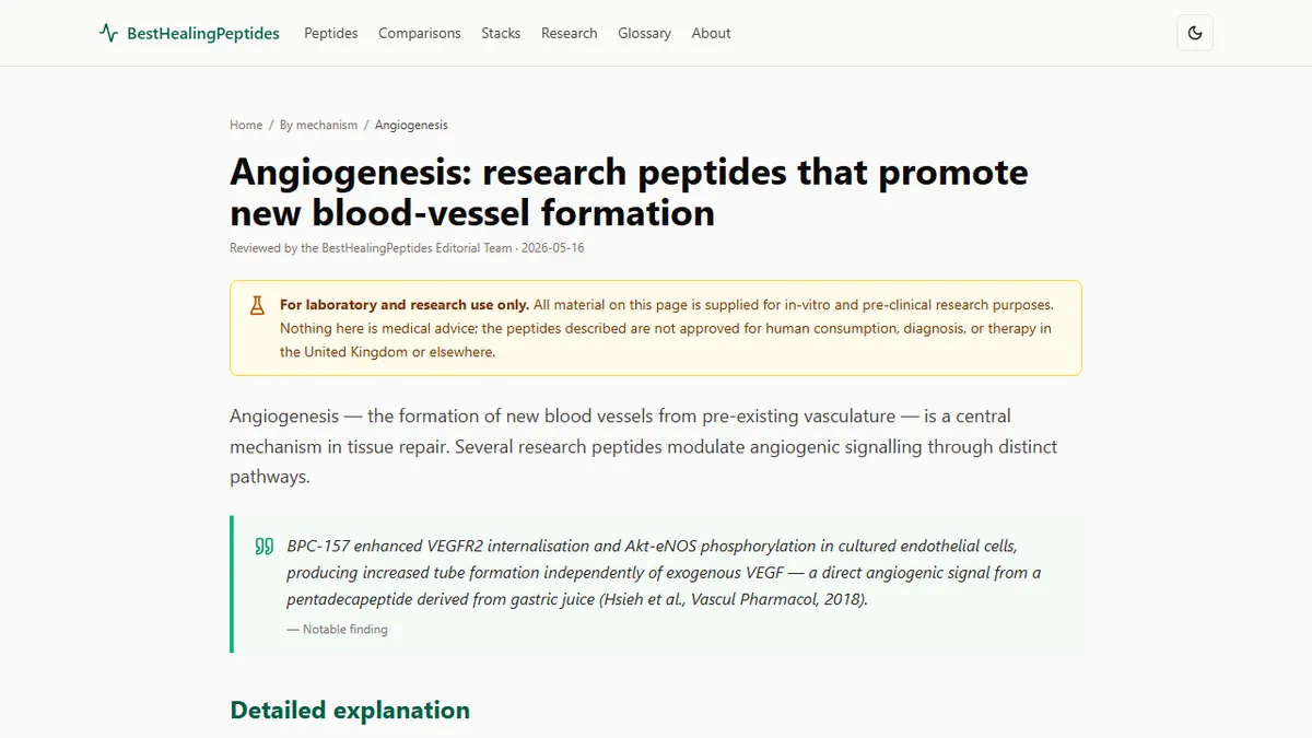
BPC-157 takes what is, from a pharmacological standpoint, an unusual route into the angiogenic cascade. Hsieh and colleagues demonstrated in endothelial cell culture experiments that BPC-157 increases VEGFR2 internalisation and downstream Akt-eNOS phosphorylation without requiring the addition of exogenous VEGF. The receptor is activated (or at minimum sensitised) in a ligand-independent manner.
The practical consequence is that BPC-157's angiogenic effect does not depend on VEGF concentration in the wound microenvironment. In ischaemic tissue beds where VEGF signalling may be impaired by hypoxia-induced receptor downregulation, or in research models where VEGF expression is deliberately suppressed as a control, BPC-157 should in principle retain angiogenic activity through this direct receptor pathway. This is sometimes described in the Sikiric-group literature as the "vessel-rescuing" effect (the observation that BPC-157 can restore microvascular patency and promote revascularisation even in conditions that compromise the canonical VEGF signal).
In injured rodent tissue, this translates to measurable increases in microvessel density within granulation tissue at tendon, muscle, and bowel anastomosis sites, alongside VEGF upregulation in the healing zone that may be a secondary amplifying signal rather than the primary angiogenic trigger.
TB-500 and Tβ4: Actin-Driven Endothelial Migration as the First Sprouting Step
Angiogenesis begins before a single new capillary has formed. The first step is endothelial tip-cell extension: individual cells at the front of an existing vessel extend filopodia into the surrounding matrix, guided by chemotactic gradients, and migrate directionally toward the angiogenic stimulus. This migration step requires cytoskeletal remodelling, specifically the rapid polymerisation of actin at the leading edge of the cell.
TB-500 (the LKKTETQ fragment) and full-length thymosin beta-4 promote this first step directly. By modulating the G-actin to F-actin ratio (maintaining a pool of monomeric G-actin available for rapid polymerisation), they enhance the capacity of endothelial cells to form lamellipodia and migrate in response to chemotactic stimuli. In full-thickness murine wound models, topical thymosin beta-4 has been shown to approximately halve wound closure time, with parallel increases in microvessel density measured by CD31 immunohistochemistry.
The TB-500 mechanism positions these peptides upstream of VEGF in the angiogenic sequence: they enhance the cellular machinery required to execute the migration response rather than increasing the strength of the pro-angiogenic signal itself. This is a meaningful mechanistic distinction when designing combination experiments with BPC-157 — the two peptides promote angiogenesis through largely non-overlapping pathways and could theoretically be additive.
LL-37: FPR2-Mediated Angiogenesis in Infected or Chronic Wound Beds
LL-37 is the only human cathelicidin, and its angiogenic properties emerge from a completely different receptor biology than any of the other peptides in this group. LL-37 engages the formyl peptide receptor 2 (FPR2, also known as FPRL1) expressed on endothelial cells. FPR2 is a G-protein-coupled receptor originally characterised in the context of resolvin and lipoxin-mediated inflammation resolution; its activation by LL-37 triggers downstream VEGF production and promotes endothelial tube formation.
The physiological context of LL-37's angiogenic activity is important for understanding when it is the appropriate experimental tool. LL-37 is released from neutrophils, keratinocytes, and macrophages at wound sites, particularly in infected or chronically inflamed wounds where antimicrobial defence is simultaneously required. Its angiogenic signal is therefore contextually embedded in the innate immune response rather than in the clean proliferative healing phase. EGFR transactivation by LL-37 also promotes keratinocyte migration, adding a re-epithelialisation component to its wound-bed activity.
For researchers designing experiments in clean, non-infected wound models in immunocompetent animals, LL-37 has the complication of also activating toll-like receptor 9 and triggering pro-inflammatory cytokine release at supraphysiological concentrations. This dual biology (pro-healing and pro-inflammatory simultaneously) means that dose selection is particularly critical in LL-37 angiogenesis experiments, and that the angiogenic signal may be confounded by concurrent inflammatory activation.
GHK-Cu: Lysyl Oxidase, Wnt/β-Catenin, and Matrix Scaffolding for New Vessels
GHK-Cu's contribution to angiogenesis operates through matrix architecture rather than direct receptor activation. New capillaries require a physical scaffold to invade: a matrix of fibronectin, laminin, and cross-linked collagen through which endothelial tip cells can extend. Lysyl oxidase, the copper-dependent enzyme that cross-links collagen and elastin fibres, is activated by GHK-Cu-mediated copper delivery to fibroblasts and smooth muscle cells. A properly cross-linked extracellular matrix provides the mechanical substrate and adhesion sites that endothelial cells require for directional sprouting and tube stabilisation.
GHK-Cu also activates Wnt/beta-catenin signalling in dermal papilla cells and possibly in perivascular fibroblasts, a pathway with established roles in tissue morphogenesis and vasculogenesis. In the context of wound healing, Wnt activation promotes the assembly of perivascular supporting cells (pericytes and smooth muscle cells) that stabilise newly formed capillaries and prevent regression once the acute angiogenic stimulus recedes. Without adequate pericyte recruitment, new vessels are fragile and prone to collapse, limiting the functional benefit of the initial angiogenic response.
Choosing the Right Angiogenic Probe for Your Research Model
The mechanistic diversity among these four peptides translates directly into a practical decision framework for research design. If the experimental question concerns direct VEGFR2 signalling in endothelial cells, or angiogenesis under conditions of VEGF limitation, BPC-157 is mechanistically aligned. If the question is about endothelial migration speed and cytoskeletal dynamics in the early sprouting phase, TB-500 or full-length Tβ4 are the appropriate tools. If the model involves infected wound beds where antimicrobial and angiogenic activities are simultaneously required, LL-37 is the only compound in this group that addresses both demands. If the question centres on matrix quality and the stability of new vasculature rather than its initial formation, GHK-Cu's matrix-scaffolding mechanism is the relevant entry point.
Using any of these peptides interchangeably on the grounds that they all "promote angiogenesis" conflates distinct molecular mechanisms and risks attributing outcomes to the wrong pathway. Designing a study with the specific upstream mechanism in mind (and selecting appropriate molecular controls such as VEGFR2 inhibitors, actin-depolymerising agents, or copper chelators) produces interpretable data that adds to mechanistic knowledge rather than merely confirming a shared downstream readout.